Hot NewsReal Cross Section Of A Bone / Cross Section Of A Bone A Cross Sectional View Of A Femur Bone Download Bone Marrow Is The Soft Highly Vascular And Flexible Connective Tissue Within Bone Cavities Which
Real Cross Section Of A Bone / Cross Section Of A Bone A Cross Sectional View Of A Femur Bone Download Bone Marrow Is The Soft Highly Vascular And Flexible Connective Tissue Within Bone Cavities Which
Real Cross Section Of A Bone / Cross Section Of A Bone A Cross Sectional View Of A Femur Bone Download Bone Marrow Is The Soft Highly Vascular And Flexible Connective Tissue Within Bone Cavities Which. Cross‐sectional area is derived from the integral of the bone mass profile across the narrow region. And why does the marrow stop where it does, and so sharply? Slides have to be made this way because the matrix of bone is too hard to be cut with a knife as the other tissues are. Cartilaginous area at the ends of long bones where lengthwise growth takes place in the immature skeleton. Compact bone is the outer layer and the spongy bone forms the.
An outer 'fibrous layer' containing mainly fibroblasts, and an inner 'cambium layer' containing progenitor cells. In addition, cortical bone thickness at anterior, posterior, medial, and lateral parts of the bone section was measured. Home » unlabelled » real cross section of a bone : This page discusses the calculation of cross section properties relevant to structural analysis, including centroid, moment of inertia, section modulus, and where da represents the area of an infinitesimally small element, a is the total area of the cross section, and x and y are the. Anyone dealing in ivory needs to know the laws regulating its sale, display and transportation.
Alveolar Bone Its Anatomical Physiological And Structural Characteristics from periobasics.com No need to register, buy now! Find the perfect bones cross section stock photo. This is the same reason why the slightest touch hurts so much. In addition, cortical bone thickness at anterior, posterior, medial, and lateral parts of the bone section was measured. It consists of two layers; Anatomy of a flat bone. Use colored pencils to draw and label the following structures as they appear using the 40x objective, or by looking at an image from the internet. Anyone dealing in ivory needs to know the laws regulating its sale, display and transportation.
Browse 53 bone marrow cross section stock photos and images available, or search for bone cross section or bone cells to find more great stock photos and pictures.
Cross section of the leg through the soleus muscle: The cell line involved in osteogenesis consists of preosteoblasts, osteoblasts, osteocytes and bone. Use colored pencils to draw and label the following structures as they appear using the 40x objective, or by looking at an image from the internet. End of a long bone. There are trabeculae in spongy bone which gives its sponge like appearance. The geometrical properties generated from the ct image included as follows: Huge collection, amazing choice, 100+ million high quality, affordable rf and rm images. Label the haversian canal, osteocyte (mature bone cell) in lacuna, and canaliculi. An outer 'fibrous layer' containing mainly fibroblasts, and an inner 'cambium layer' containing progenitor cells. Compact bone is the outer layer and the spongy bone forms the. Two types of bone tissues in cross section of a long bone : Related posts of cross section of a long bone bone structure right foot. This page discusses the calculation of cross section properties relevant to structural analysis, including centroid, moment of inertia, section modulus, and where da represents the area of an infinitesimally small element, a is the total area of the cross section, and x and y are the.
This is known as the periosteum. There are trabeculae in spongy bone which gives its sponge like appearance. Find the perfect bones cross section stock photo. This file is licensed under the creative commons attribution 3.0 unported license.: The cortical bone equivalent area of the cross‐section of the region of interest (femoral neck or shaft), with all soft tissue voids (trabecular and cellular spaces) eliminated (cm 2).
Schematic Diagram Of Long Bone Cross Section 47 Download Scientific Diagram from www.researchgate.net The cortical bone equivalent area of the cross‐section of the region of interest (femoral neck or shaft), with all soft tissue voids (trabecular and cellular spaces) eliminated (cm 2). Slides have to be made this way because the matrix of bone is too hard to be cut with a knife as the other tissues are. Muscles and bones of the human body 12 photos of the muscles and bones of the human body anatomy bones of the human body quiz, major muscles and bones in the human body, muscles and bones in the human body, number of muscles and bones in the human body. I don't find it enhances the image. Body size standardization was done, using the following equations: Bone is a dynamic biological tissue, composed of various metabolically active cells that are integrated into a rigid framework. Each bone in your body is made up of three main types of bone material: Related posts of cross section of a long bone bone structure right foot.
The cell line involved in osteogenesis consists of preosteoblasts, osteoblasts, osteocytes and bone.
Generally speaking, it is very easy to recognize a cross section through the leg, mostly due to the tibia. I don't find it enhances the image. Human bone, cross section diagram of femur showing osteon, veins, marrow. This is the same reason why the slightest touch hurts so much. This bone is located directly beneath the skin on the anterior aspect of the leg (top of the image). The cell line involved in osteogenesis consists of preosteoblasts, osteoblasts, osteocytes and bone. An outer 'fibrous layer' containing mainly fibroblasts, and an inner 'cambium layer' containing progenitor cells. Find the perfect bones cross section stock photo. Browse 53 bone marrow cross section stock photos and images available, or search for bone cross section or bone cells to find more great stock photos and pictures. Cross section of long bone. Related posts of cross section of human bone diagram muscles and bones of the human body. Label the haversian canal, osteocyte (mature bone cell) in lacuna, and canaliculi. Use colored pencils to draw and label the following structures as they appear using the 40x objective, or by looking at an image from the internet.
It consists of two layers; Each bone in your body is made up of three main types of bone material: Why is the marrow red? Would it be a good thing to show the epiphyseal plate? Related posts of cross section of human bone diagram muscles and bones of the human body.
Cross Section View Of A Human Femur Bone Showing Trabecula Stock Photo Alamy from c8.alamy.com Find the perfect bones cross section stock photo. Huge collection, amazing choice, 100+ million high quality, affordable rf and rm images. Label the haversian canal, osteocyte (mature bone cell) in lacuna, and canaliculi. You may do so in any reasonable manner, but not in any way. Human bone, cross section diagram of femur showing osteon, veins, marrow. Cross section of the leg through the soleus muscle: Muscles and bones of the human body 12 photos of the muscles and bones of the human body anatomy bones of the human body quiz, major muscles and bones in the human body, muscles and bones in the human body, number of muscles and bones in the human body. Would it be a good thing to show the epiphyseal plate?
It forms the hard exterior (cortex) of bones.
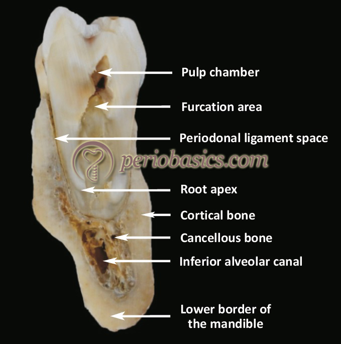
This page discusses the calculation of cross section properties relevant to structural analysis, including centroid, moment of inertia, section modulus, and where da represents the area of an infinitesimally small element, a is the total area of the cross section, and x and y are the. Cross section of mandible at first molar region showing cortical and spongy bone basic concepts in osteogenesis. Terms in this set (3) epiphysis. Compact bone is the outer layer and the spongy bone forms the inner layer. (area/long bone length 3) ∗ 10 8. Spongy bone and compact bone. This bone is located directly beneath the skin on the anterior aspect of the leg (top of the image). Two types of bone tissues in cross section of a long bone : Compact bone, spongy bone, and bone marrow. Anyone dealing in ivory needs to know the laws regulating its sale, display and transportation. It consists of two layers; Then, fill in the table below to describe each. Translucence is an ivory characteristic that can be helpful in differentiating it from bone, as bone is an opaque substance.
The compact bone is made up of osteon cross section of a bone. The geometrical properties generated from the ct image included as follows:


0 comments